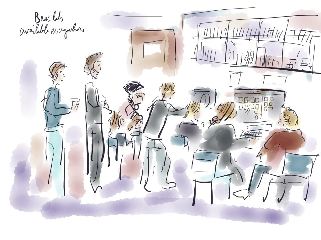
Today, Thomas Picht and Lucius Fekonja were teaching a group of seven students how to prepare a brain surgery. All of it happens on Brainlab, a software that is considered the “Apple” among the medical software, says Thomas.

The software is available from any computer in the hospital. The data of the MRI appears directly in the program. First part is to find the patient and access to the relevant images.

The next step is a fusion of the images that are available.

The scan appears in three dimensions, according to the series of images generated by the MRI. From those 3 series, the software generates a 3D visualisation, on the up-left corner.

The next step is to “draw” the tumor of the patient. Several tools exists in the program. On one of the three 2D images, the student “draws” the tumor by manually highlighting the borders of that slightly zone. An algorithm in the software guides this drawing by coloring automatically the tumor. The “drawing” appears as one “slice” of the tumor in the 3D representation.

After having done the same work on another 2D, the tumor appears now in 3D. the student drawing on a second of the 2D images, the tumor now appears as a 3D representation in the viewer. This has to be refined now, and depending on the clarity of the definition of the tumor, the manual result can show a sensible difference with the result of the algorithm. Thomas explains that even very experienced users can obtain slightly different drawings: the challenge is that the border is never completely sure. They discuss now about how to access that tumor, there are several possibilities with various difficulties.

What can we do after this, asks Thomas? “Tractography!” reply the students. They have been learning the day before from Lucius about that relatively new technique of visualization of the most important networks of the white matter: those related to motric and speech functions. Those “circuits” are statistically computed from MRI data (that’s for another post). Most importantly, drawing those tracks enable the surgeon to avoid damaging them during the operation.

This track, in blue, was highlighted successfully by the student. “When the track is so neatly appearing on the visualization software, you can be sure that it’s an important one”, says Thomas.
That was it for the day. Last discussion, Thomas explains about their current research interest in more advanced modes of data visualisation. Was the software clear enough? Do we need an hologram to visualise the objects in 3D? The opinions are divergent. But all seem very satisfied by this thourough hands-on introduction. Thomas conclude: “The surgeons do exactly the same that you did today. They only do it a bit faster!”